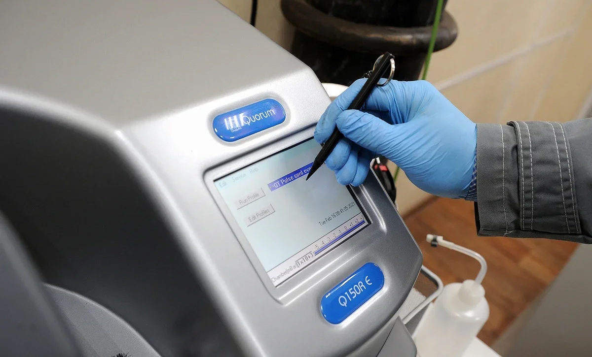
#ARCTIC. #SIBERIA. THIS IS TAIMYR. The first in Russia TESCAN TIMA system – an electron microscope for automatic mineralogical analysis was launched at the Engineering Support Center (ESC) of Nornickel Polar division. The analytical complex was assembled by the company’s order and can determine about 200 minerals.
“The microscope sees a black and white picture, it has no concept of color rendering. We can say there are 2000 shades of gray here. The so-called reflected electrons (BSE), the signal intensity of which is sensitive to the average atomic number of the material (and therefore to its density). Due to this feature, grains of different minerals tend to have different shades of gray in BSE images. The samples contain various minerals: our sulphides and silicates. They appear darker in images. And light ones are metal-containing sulphides or oxides”, says Vladimir Velichko, leading engineer of the Polar division engineering support center’s material and chemical analysis laboratory.

For analysis, the ore sample is reduced, filled with epoxy resin and allowed to solidify. It takes 16 hours. First, the microscope divides the surface of the sample into sectors and takes pictures. How long it takes depends on the ore composition. The resulting ‘washer’ is polished and covered with a thin layer of carbon. The microscope cassette roll can hold up to seven of these samples. If you need photographs of samples or just need to view them in an enlarged format, it is enough to choose raw materials with a smoother surface. Complicated preparation, as in the first case, is not required.
Specialists use gloves because organic compounds cannot be placed in the microscope. They will settle on the detectors, and this can lead to incorrect results.

The microscope examines ore samples by touch. Scanning electron microscopy methods are based on the interaction of a finely focused electron beam with the sample surface. Then the detectors collect different types of response signals, the so-called reflected electrons, bremsstrahlung and characteristic radiation, secondary electrons. The program gives the operator a black and white picture. The microscope itself colors the images, because it already knows the name and color of each mineral.
The system does both chemical and phase analysis of samples, which is very important for ore cleaning. It is possible to find out both the amount of minerals and what metals they carry. Thanks to these data, the processing of mineral raw materials, the method of extraction and crushing are adjusted. Previously, mineralogical analysis in the laboratory was done using optical microscopes.
“Optical mineralogical analysis is complex and time consuming. Lab technicians could sit and look through a microscope all day to identify and describe minerals. One sample could be studied for a week”, says Sergey Bolshakov, head of the laboratory for material and chemical analysis of the engineering support center.
The analysis of one sample takes, depending on the task, from 20 minutes to one and a half days. The flow of samples to the ESC laboratory is continuous. The specialists have not yet finished ‘training’ the system. New tasks appear every day. There are minerals that have not yet been recognized, perhaps these are some new, undiscovered compounds.
So far, such a device is the only one in Russia. It was assembled by the special order of Nornickel. Representatives of other domestic enterprises regularly call up the ESC employees to find out how effective the system is, how it has shown itself. The Polar division specialists note: everything works at its best.
Text: Vera Karpenko, Angelica Stepanova, Photo: Nikolay Shchipko


